It's important to us to keep you informed about changes in medical physics regulations -- and it's also our pleasure to keep you informed as part of our commitment to customer service.
Read our blog often for the latest updates and expert medical physics advice from our skilled team.
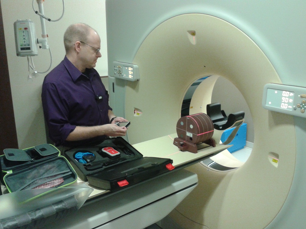
What Can We Say About Patient Dose in CT Exams?
Posted on Tuesday, April 2nd, 2013, under CT
By Stephen E. Hale Jr., Ph.D.
Radiation dose from Computed Tomography (CT) exams has recently become a hot topic among the medical community, the legal community, and the public in general. Overdoses from brain perfusion studies at several medical facilities in California and Alabama have contributed to this rise in concern. But what can a CT technologist or radiologist actually tell their patients about the amount of radiation administered during a CT exam?
Each CT machine will typically provide both an estimated CTDIvol and DLP value before an exam is conducted. CTDIvol stands for volume Computed Tomography Dose Index, while DLP stands for Dose-Length Product. Neither of these is actually a measure of the dose a patient will get from the exam. They are actually designed as metrics for comparing one protocol to another, one machine to another, or even one facility to another, in terms of the amount of radiation produced by the system. As such, they can be used to guide adjustments to the techniques of a given protocol, such as kVp, mA, rotation time, mAs, or even pitch for helical scans.
Dose is defined as the amount of energy absorbed from radiation passing through material divided by the amount of mass that is actually absorbing the energy. The more energy absorbed in the same amount of matter, such as a patient’s body, the more dose is absorbed, and therefore the greater potential for damage. Similarly, the same amount of energy absorbed in a smaller body will also result in a greater potential for damage.
The CT machine has no knowledge about the patient who will be scanned nor what portion of the patient’s anatomy will be examined. If a large patient is scanned with the same techniques as a small patient, the smaller patient will have less mass exposed to the same amount of radiation, and thus experience a higher dose. Different portions of our bodies are more sensitive to radiation than others. Thus having a CT exam with a set of technique factors over a patient’s feet will have much less detriment to the patient than the same exam over a patient’s head.
A good analogy to this situation is that of a tachometer in a car relative to the car’s speed. The tachometer displays how many revolutions per minute the engine is spinning the crankshaft, but it has no knowledge about the gear selected by the transmission or the size of the wheels and tires on the car. For a given RPM, a higher gear means a faster speed. Similarly, if the wheels and tires are replaced with a larger set, a given RPM of the engine and the same gear will result in a faster speed. The CTDIvol and DLP values are the equivalent of the tachometer, reporting how the CT machine is performing. But without information about the patient undergoing the procedure, nothing can be said about the dose absorbed.
Without knowledge of the patient’s size or the anatomy being scanned by the CT, it is not possible to tell the patient how their radiation dose compares to other sources or exams. With images of the patients and information about the parameters of the exam on the CT system, physicists can calculate actual radiation doses to patients. This is one of the many services provided by Integrated Science Support, Inc.
References:
- “Two more hospitals report CT scan radiation overdoses” LA Times, Online 8/3/2010
Radiographic Technique Still Matters For Image Quality and Patient Dose
Posted on Saturday, March 23rd, 2013, under Radiography

Shirley Bartley, M.B.A., RT (R)(N)
The new technologist at the hospital radiology department went to do a portable in the ICU. The patient looked average size. She looked for the technique chart to set the correct exposure factors. There was no chart to be found. She guessed at the technique based on what she could remember from the last place she worked. When the new tech returned to the department the supervisor was reviewing the image. “You are going to have to repeat this portable chest. There is an image quality problem. You didn’t penetrate the mediastinum.” The supervisor told the new tech.
If the kVp is too low the anatomy will not be properly penetrated. The fine detail in dense areas of the body will just not be there. This will jeopardize the radiologist ability to make a correct interpretation of the patient’s condition.
A few minutes later the new tech was working in room 2 with a seasoned employee. The new tech selected the radiographic technique from the anatomic programing feature on the operator’s consol. “Don’t use that. It doesn’t work. I have my own technique.” The new technologist made a mental note to try to remember the technique.
Without standard technique systems that are used by everyone image quality will be inconsistent. It is difficult for the radiologist to see changes in the patient’s condition when totally different radiographic technique is used. Technologist that just “remember” their own techniques are frequently wrong.
After lunch the new tech was working with a radiology student. They did an abdomen using automatic exposure control (AEC). Viewing the image, the new tech pointed out that the exposure indicator value was well above the acceptable range. The students said, “They don’t pay any attention to the number here.”
With digital systems the image that is over exposed no longer comes out black. The only way we can determine that the patient was over exposed is with the exposure indicator value. The AEC unit that is not properly calibrated will not produce the correct quantity of radiation for the image. When the exposure indicator value is ignored the patients may be overexposed unnecessarily.
These three situations describe common problems for image quality and patient dose. ISS, Inc. can assist you in maximizing image quality while keeping patient dose as low as reasonably achievable (ALARA). For more information access our recent white paper.